We use cookies to make your experience better. To comply with the new e-Privacy directive, we need to ask for your consent to set the cookies. Learn more.
Vitrification - A front-runner of human oocyte and embryo cryopreservation technology
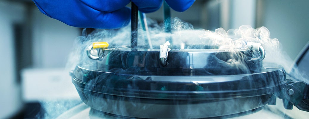
Cryopreservation involves the cooling of living cells and tissues down to minus 196°C to arrest all biological activities and preserve them for future use. In assisted reproductive technology (ART), the preservation of both sperm and oocytes (gametes) as well as embryos is a medically recognized treatment option for patients at risk of losing their fertility due to aggressive medical treatments, reproductive disorders, or as a part of the aging process.
According to the Centers for Disease Control and Prevention (CDC), approximately 1.6% of infants born every year in the US are conceived using ART, including in vitro fertilization (IVF). To maximize the efficiency of IVF with every gamete retrieved, the field of reproductive cryobiology has been devoted to developing methods for successfully cryopreserving gametes and their resulting embryos. It is worth giving a brief review of gametogenesis and the IVF process at this time (included below) to help truly grasp and appreciate the significance of successful cryopreservation.
Human females are born with a lifetime supply of primary oocytes arrested at prophase I prior to the first meiotic division. The process of meiosis I resumes at puberty and completes maturation forming the secondary oocyte. Ovulation occurs approximately once a month with each ovary alternating in the release of a mature metaphase II (MII) oocyte to the fallopian tube where it has the opportunity to be fertilized and receive another set of 23 chromosomes as it travels down toward the uterus. After ovulation, the oocyte’s nuclear and cytoplasmic maturation continues providing a 12 to 24 hr window for successful fertilization. Once fertilized, the activated oocyte resumes the rest of meiosis and finally completes the first mitotic cell division resulting in a 2-cell embryo.
The human male reproductive system differs from females in that primordial germ cells in mature testes divide to form spermatogonia and in turn generate primary spermatocytes through mitosis. Subsequently, each primary spermatocyte moves into the seminiferous tubules where it duplicates its DNA content and undergoes meiosis I and II. This gives rise to four haploid spermatids, ultimately contributing to millions of sperm in a single ejaculation event. During division, a subset of spermatogonia remains undivided to prevent depletion allowing for the steady replenishment of primary spermatocytes through mitotic cell division.
A diagnosis of infertility is given to couples who have been trying to conceive for 12 months or more without success1. At fertility clinics, an IVF cycle includes extracting gametes, fertilizing the mature MII oocytes in vitro by conventional or intracytoplasmic sperm injection (ICSI), and transferring the resulting embryos back to the mother’s uterus. IVF patients undergo controlled ovarian hyperstimulation prior to ultrasound directed follicular aspiration (UDFA) in order to harvest mature MII oocytes. Then one (or two) viable high quality embryo that underwent Preimplantation Genetic Diagnosis (PGD) is selected for transfer. If there is a surplus of good quality embryos, they are cryopreserved for future use.
Gamete quality plays an essential role in successful IVF assisted pregnancies. The earliest success in human sperm retrieval after cryopreservation was documented in 19492; however, successful pregnancies with cryopreserved oocytes was not reported until 19863. Since then, nearly two decades have been dedicated toward optimizing techniques for safely cryopreserving human oocytes and surplus embryos. The first successful pregnancy utilizing cryopreserved human cleavage-stage embryos and oocytes3-5 established the slow-freezing approach as the primary technique for cryopreservation. However, inconsistency in outcome, time consuming and slow cooling steps (ranging from 2 to 5 hrs), cytotoxicity of cryoprotectants (CPAs), and most importantly damage due to the intracellular crystallization of water presents concerns and challenges to the use of slow-freezing for ART.
Another methodology utilized for cryopreserving gametes and embryos is an ice-free process known as vitrification which has been successfully demonstrated through numerous successful pregnancies6-8. Vitrification (glass-like solidification) is accomplished by rapid cooling with increased viscosity and a depressed freezing point with aid of CPAs9. Although the ice-free cryopreservation of mouse embryos was first reported in 198510, vitrification was not seriously considered as an alternative to the slow-freezing method due to the cytotoxic effects associated with high concentrations of CPAs (3 to 4 fold higher than the slow-freezing method). The necessity of using high concentrations was thought to cause higher intracellular concentrations of CPA and presented challenges for its use in the IVF field. While the vitrification method was shown to be effective for all developmental stages of human embryo4,6,7, it was not until 2005 when the Kuwayama group successfully demonstrated the vitrification of human oocytes using a cryodevice known as Cryotop to reduce the volume of vitrification medium to below 0.1 µL on the surface of a fine polypropylene strip for rapid cooling and warming rates11, to be fully accepted as an effective method. Notably, in a recent study, only a fraction of permeable CPAs were found in vitrified mouse zygotes compared to the slow freezing method12, possibly due to a rapid cooling and thawing process eliminating a prolonged exposure to CPA containing solutions. Irvine Scientific launched the first FDA cleared vitrification product in 2006. Currently, vitrification enjoys wide acceptance in the ART field and is becoming a frontrunner for cryopreservation approaches.
The successful utilization of vitrification for IVF requires a balance of three major properties: 1) CPA concentration, 2) rapid cooling and warming, and 3) media volume (Fig.1) to prevent the intracellular crystallization of water.
|
|
 |

Vitrification solutions contain both permeable and non-permeable CPAs (Table 1) to increase viscosity and lower the freezing point of the solutions. Optimal concentrations of permeable CPAs depend on the kinetics of cooling and warming steps. Furthermore, limited volume of vitrification medium on the cryodevice helps facilitate rapid cooling and warming.
The following example utilizing FUJIFILM Irvine Scientific’s Vit Kit has been included to give greater detail regarding the steps and processes involved. When the embryo is placed in equilibration solution (ES), the intracellular water is osmotically replaced with DMSO/EG (Fig. 2, ES), and the system reaches equilibrium.

Figure 2: Schematic presentation of vitrification and warming procedure. During vitrification step (green arrows), an embryo is exposed to Equilibration Solution (ES) containing DMSO/EG (CPAperm) from 6 to 10 min and undergoes osmotic change (hypertonic → isotonic state). When osmotic pressure reaches equilibrium, the embryo is placed in Vitrification Solution (VS) containing higher concentrations of DMSO/EG and 0.5M sucrose (CPAnon-perm) for 30 seconds to dehydrate, loaded onto a cryodevice with a very small volume of vitrification medium, and plunged into liquid nitrogen within 80 seconds. The whole vitrification step takes about 10 minutes. In the warming steps (tan arrows), the embryo is rapidly warmed in Thawing Solution (TS) for 1 minute at 37°C and undergo further dehydration. Stepwise reduction of sucrose concentrations from 1M in TS to 0.5M in Dilution Solution (DS) induces rehydration of the cells while providing an osmotic buffer. During the incubation in Wash Solution (WS, 4 min twice), the embryo is pre-equilibrated before transferring to culture medium. Inner circle: blastocyst, outer circle: zona pellucida.
Enhanced cellular dehydration is then achieved with non-permeable sucrose creating an osmotic gradient to facilitate cell dehydration (Fig. 2, VS). DMSO/EG diffuses into the cells protecting the cell membrane during rapid cooling by modifying the state of the phospholipid bilayer from fluid to partially solid13. It also reduces intracellular crystallization of water by lowering the freezing point of the solution. During the warming process, stepwise reduction of sucrose concentrations in thawing and dilution solutions (Fig.2, TS & DS) induces removal of intracellular DMSO/EG and rehydrates the cells while providing an osmotic buffer to reduce osmotic shock14 caused by the swelling of cells from the rapid influx of water. Washing solution (WS) is then utilized to equilibrate the embryo in preparation for transfer into the culture medium.

Figure 3: Post-thaw images of vitrified mouse blastocysts. Mouse Pou5f1-GFP embryos were used to evaluate morphology and POU5F1-GFP protein expression (green) after vitrification/thawing . a. Representative fluorescent images of non-vitrified Pou5f1-GFP blastocysts at 96hrs showing localized expression of POU5F1-GFP in the ICM (blue arrows). Pou5f1-GFP blastocysts were vitrified/thawed with b. Irvine Scientific Vit Kit–Freeze (PN 90133) and Vit Kit–Thaw (PN 90137) or c. a set of prototype vitrification and warming solutions. Pou5f1-GFP blastocysts vitrified/thawed in the IS media advanced to the hatched stage after 24hrs post-thaw and maintained POU5F1-GFP expression localized in the ICM. One hatched embryo (yellow arrow) showing reduced POU5F1-GFP expression. Blastocoel of a live hatching blastocyst vitrified/thawed in the prototype set remained collapsed at 24hr post-thaw (white arrows). All Images were taken with 40x objective. Top: POU5F1-GFP; Bottom: phase contrast.
Survival of vitrified/thawed embryos is often difficult to evaluate since morphological observation alone would not definitively predict developmental ability and even genomic integrity of the embryos. We investigated this concern by employing mouse embryos expressing green fluorescent protein under control of Oct4 (also known as Pou5f1) regulatory elements15 and evaluating morphology and POU5F1-GFP expression after vitrification/thawing (Fig.3). POU5F1-GFP expression was used as a molecular marker to determine the impact of vitrification on the embryos and their genomic integrity. Pou5f1-GFP embryos were cultured for 96hrs and vitrified with IS Vit Kit–Freeze (PN 90133), and plunged into liquid nitrogen after being loaded onto a device known as Cryolock which is made from a single material to ensure an enhanced temperature coefficient. The embryos were subsequently warmed with Vit Kit–Thaw (PN 90137) and rehydrated. The vitrified/thawed Pou5f1-GFP embryos showed expansion of blastocoels at 2 hr post-thaw (Fig.3, b). Pou5f1-GFP embryos also advanced to the hatched stage at 24 hr post-thaw and maintained POU5F1-GFP expression localized in the ICM, indicating sustained biological activity and intact genomic function. One aged hatched embryo showed reduced POU5F1-GFP expression as a possible sign of degradation (Fig. 3b, yellow arrow). Pou5f1-GFP embryos were also vitrified with a set of prototype solutions containing different basal medium known to effectively culture human embryos to test the role of culture medium in vitrification and thawing. We observed expansion of blastocoels at 2hr post-thaw (Fig.3, c) and consistent survival rates at 2 hr and 24 hr post-thaw. The blastocoel of one live hatching blastocyst remained collapsed at 24 hr post-thaw (white arrows) while expression of POU5F1-GFP was maintained in the ICM. This could potentially be due to insufficient equilibration during the warming process. The change in basal medium seemed to have less impact on the integrity and biological activities of embryos after thaw. Utilizing POU5F1-GFP as a biomarker, we visualized and demonstrated the safety of vitrification for cryopreservation. More specifically we showed vitrification’s ability to: 1) arrest all biological activities and reactivate after thaw, 2) preserve morphological and genomic integrity, 3) and to maintain developmental competency after thaw.
Cryopreservation of sperm and oocytes is an essential technology meeting the difficulties and setbacks oftentimes associated with infertility. It provides patients with greater options for preserving their fertility, including situations that require medical treatments that may negatively affect reproductive functions. It is noteworthy that promising outcomes of first IVF cycles using vitrified oocytes have been accumulating8,16,17 with some IVF clinics offering selected patients a “freeze-all” cycle18-20 to cryopreserve all retrieved oocytes as an option to treat infertility. Though room for improvement is continuously needed, these reports validate vitrification as a reliable and fast growing clinical application with great consistency and better outcomes.
Vit Kit is a registered trademark of FUJIFILM Irvine Scientific. All other trademarks are the property of their respective owners.
References:
- Beers, M. H. The Merck Manual of Diagnosis adn Therapy, 18th Edition. (Merck, 2006).
- Polge, C., Smith, A. U. & Parkes, A. S. Revival of spermatozoa after vitrification and dehydration at low temperatures. Nature 164, 666 (1949).
- Chen, C. Pregnancy after human oocyte cryopreservation. Lancet 1, 884-886 (1986).
- Trounson, A. & Mohr, L. Human pregnancy following cryopreservation, thawing and transfer of an eight-cell embryo. Nature 305, 707-709 (1983).
- Zeilmaker, G. H., Alberda, A. T., van Gent, I., Rijkmans, C. M. & Drogendijk, A. C. Two pregnancies following transfer of intact frozen-thawed embryos. Fertil Steril 42, 293-296 (1984).
- Wennerholm, U. B. et al. Children born after cryopreservation of embryos or oocytes: a systematic review of outcome data. Hum Reprod 24, 2158-2172, doi:10.1093/humrep/dep125 (2009).
- Liebermann, J. & Tucker, M. J. Comparison of vitrification and conventional cryopreservation of day 5 and day 6 blastocysts during clinical application. Fertil Steril 86, 20-26, doi:10.1016/j.fertnstert.2006.01.029 (2006).
- Nagy, Z. P., Anderson, R. E., Feinberg, E. C., Hayward, B. & Mahony, M. C. The Human Oocyte Preservation Experience (HOPE) Registry: evaluation of cryopreservation techniques and oocyte source on outcomes. Reprod Biol Endocrinol 15, 10, doi:10.1186/s12958-017-0228-7 (2017).
- Fahy, G. M., MacFarlane, D. R., Angell, C. A. & Meryman, H. T. Vitrification as an approach to cryopreservation. Cryobiology 21, 407-426 (1984).
- Rall, W. F. & Fahy, G. M. Ice-free cryopreservation of mouse embryos at -196 degrees C by vitrification. Nature 313, 573-575 (1985).
- Kuwayama, M., Vajta, G., Kato, O. & Leibo, S. P. Highly efficient vitrification method for cryopreservation of human oocytes. Reprod Biomed Online 11, 300-308 (2005).
- Vanderzwalmen, P. et al. Lower intracellular concentration of cryoprotectants after vitrification than after slow freezing despite exposure to higher concentration of cryoprotectant solutions. Hum Reprod 28, 2101-2110, doi:10.1093/humrep/det107 (2013).
- Zaca, C. & Borini, A. Chapter 8 Human Oocytes Slow-Rate Freezing: Methodology. Methods Mol Biol 1568, 105-117, doi:10.1007/978-1-4939-6828-2_8 (2017).
- Liebermann, J. Chapter 11 Human Embryo Vitrification. Methods Mol Biol 1568, 141-159, doi:10.1007/978-1-4939-6828-2_11 (2017).
- Gilbert, R. S., Nunez, B., Sakurai, K., Fielder, T. & Ni, H. T. Genetic mouse embryo assay: improving performance and quality testing for assisted reproductive technology (ART) with a functional bioassay. Reprod Biol Endocrinol 14, 13, doi:10.1186/s12958-016-0149-x (2016).
- Yoon, T. K. et al. Pregnancy and delivery of healthy infants developed from vitrified oocytes in a stimulated in vitro fertilization-embryo transfer program. Fertil Steril 74, 180-181 (2000).
- Kuleshova, L., Gianaroli, L., Magli, C., Ferraretti, A. & Trounson, A. Birth following vitrification of a small number of human oocytes: case report. Hum Reprod 14, 3077-3079 (1999).
- Surrey, E. et al. Freeze-all: enhanced outcomes with cryopreservation at the blastocyst stage versus pronuclear stage using slow-freeze techniques. Reprod Biomed Online 21, 411-417, doi:10.1016/j.rbmo.2010.04.008 (2010).
- Toftager, M. et al. Cumulative live birth rates after one ART cycle including all subsequent frozen-thaw cycles in 1050 women: secondary outcome of an RCT comparing GnRH-antagonist and GnRH-agonist protocols. Hum Reprod 32, 556-567, doi:10.1093/humrep/dew358 (2017).
- Blockeel, C., Drakopoulos, P., Santos-Ribeiro, S., Polyzos, N. P. & Tournaye, H. A fresh look at the freeze-all protocol: a SWOT analysis. Hum Reprod 31, 491-497, doi:10.1093/humrep/dev339 (2016).

Recent Posts
-
 Meet the Team: Interview with Jennifer Brooks, Environmental Health and Safety (EHS) Manager
 -
 Meet the Team: Interview with Chandana Sharma, Senior Director, Life Sciences
 -
 Interview: Jim O’Brien is ALL IN on Customer Service
 -
 Meet the Team: Interview with Barbara Davis, R&D Manager, Assisted Reproductive Technology
 -
 Interview: How FUJIFILM Irvine Scientific is ALL IN on the Future